


In a continuously expanding field, a single principle has remained consistent to date: all genetic information originates from DNA*, a concept known as the central dogma. Fundamentally, the entirety of the reader’s being is defined by approximately 3 billion sequences of only 4 genetic building blocks*, which we call DNA. In fact, all living things are united by this system of genetic coding.
However, on April 16th of 2026, a revolutionary paper titled “Protein-templated synthesis of dinucleotide repeat DNA by an antiphage reverse transcriptase” placed the central dogma into question with its discovery of a protein with the ability to make DNA from scratch. This protein was found in over 20 different bacterial species and was observed in E. coli*, a common model bacterium*. The research was led by Alex Gao and his team at Gao Labs; they determined that the protein is a defence-associated reverse transcriptase (DRT)*, a molecular machine* only found in prokaryotic cells*.
What are DRTs and what do they do?
Reverse transcriptases are the enzymes* used to synthesize complementary strands of DNA from RNA* strands and are found across all kingdoms of life. Defense-associated reverse transcriptases (DRTs), however, are a specific class of reverse transcriptases used only by prokaryotic cells as a defense mechanism against bacteriophages*. Only 9 types of DRTs have been established, which all act as alarms or weapons, yet all have unique mechanisms of defending against phages. The newly discovered DRT3, however, is the sole enzyme in the family to show DNA synthesis “in the complete absence of a nucleic acid template” Deng, et al..
What is DRT3 and what does it look like?
DRT3 is assembled from 3 different units: DRT3a, DRT3b, and non-coding RNA (ncRNA)*. DRT3a and DRT3b are actually two distinct types of reverse transcriptases, working together. The DRT3 hexamer* complex is composed of six DRT3a units, six DRT3b units, and six strands of ncRNA.
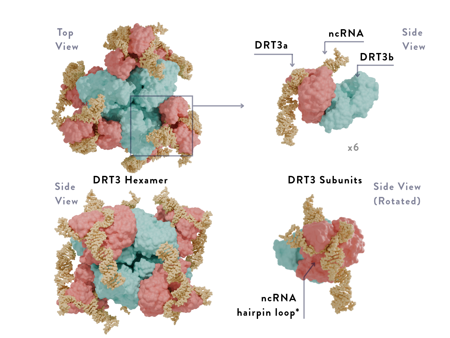
It is a beautifully symmetric molecular machine where both subunits work simultaneously, yet differently from one another, to form DNA. The claim is that DRT3 synthesizes DNA without a base copy; however, the enzyme still utilizes ncRNA. Both DTR3a and DTR3b form their own strand of DNA, except that only one has its own unique way of making DNA never observed before.
How DRT3a and DRT3b work
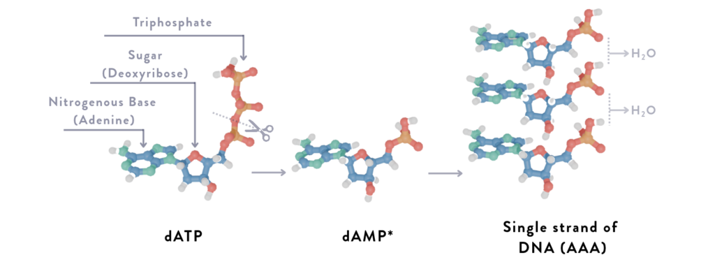
Within the bacteria’s cytoplasm* are molecules called dATP* and dCTP*, which are raw materials used for nucleotides*. DRT3 captures these floating molecules and cuts off two of the three phosphates, leaving a sugar*, a phosphate group*, and a nitrogenous base*; the remaining product is exactly what is required for DNA synthesis.
Both strands of DNA are made independently of each other. DRT3a follows the familiar procedure; it uses the ncRNA consisting of an ACACAC motif as a base, producing a repeating TGTGTG pattern called a poly(GT) strand.
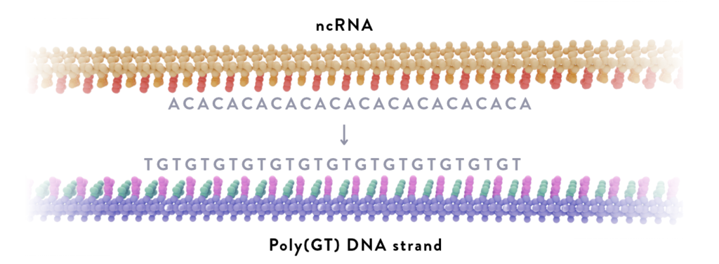
DRT3b, on the other hand, uses its own molecular geometry to form a repeating ACACAC strand, poly(AC), to complement the poly(GT) strand. DRT3b primarily uses two amino acids in its structure to bind: Glutamate (Glu26)* and arginine (Arg253)*, which act as gates that alternately allow dAMP* and dCMP* to bind together as a strand.
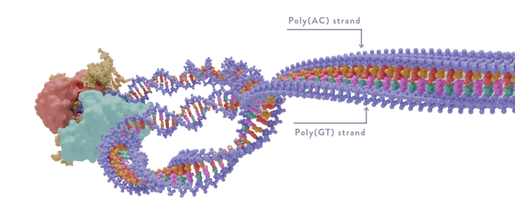
When dATP is within the active site* of DRT3b, Glu26 binds to the incoming molecule, appending dAMP to start the DNA strand. Subsequently, this alters the shape of the active site as the DNA chain moves forward, moving Glu26 out of the site and Arg253 in, allowing capture of dCTP to follow. This, in turn, pushes Arg253 back and Glu26 forward, resulting in the alternating ACACAC strand.
As both strands are pushed out of the hexamer, they both spontaneously zip together, resulting in a brand new strand of DNA consisting of complementary poly(AC) and poly(GT) strands. At first glance, the generated DNA sequence seems overly simple for such a novel discovery; however, the unique mechanism may be linked to the strand’s speculated function.
Why all the fuss for a simple sequence?
DRT3 has been shown to produce these repetitive strands of DNA in the presence of a particular protein released by phages, called ST61*. The presence of this protein is an apparent sign of a phage invasion, as it is exclusively produced by viral bacteriophages, such as T1 phages*, which were used to observe DRT3 behavior. ST61 was seen to bind to the DRT3 hexamer, kickstarting the enzyme into producing DNA.
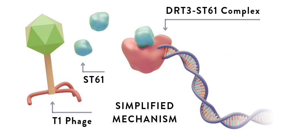
The existence of DRT3 highlights the complexity of bacteria to persevere against viruses, demonstrating a dormant molecular machine capable of fending against bacteriophage infection once initiated. While the exact function of the repetitive DNA is still under investigation, its necessity to bypass canonical DNA synthesis for immediate production may suggest that the strands act as deterrents to phage replication. It is described as potentially having a function similar to that proposed for DRT9: producing decoy DNA to bind to the DNA-binding proteins released by bacteriophages, which they use to hijack the bacteria’s genetic code to make copies.
DNA: Deoxyribonucleic acid, molecules that store all genetic information.
4 genetic building blocks: referencing the 4 bases that are the coding sequences in DNA: adenine(A), cytosine(C), thymine(T), and guanine(G).
E.coli: Escherichia coli; it’s a common bacterium that is frequently harmless and aids with digestion.
Model bacterium: Bacteria used by labs to study cellular phenomena, usually picked for being simple to grow, having high reproduction rates, and a well-studied genome.
DRT: Defense-associated reverse transcriptases; enzymes that make DNA and are specifically used to defend the cell against viral infections.
Molecular machine: An atomic or molecular assembly that uses energy to perform mechanical functions.
Prokaryotic cells: The kingdom of life comprising single-celled organisms, such as bacteria and archaea.
Enzymes: A type of protein specializing in catalyzing chemical reactions.
RNA: Ribonucleic acid, molecules that translate DNA instructions into proteins. It has various types according to their function, such as mRNA, and instead of thymine(T), it contains Uracil(U) as its pair to adenine(A).
Bacteriophage/phages: A virus that infects bacteria, replicating by hijacking the cell’s DNA to instruct its machinery to make more copies of itself.
ncRNA: non-coding RNA, RNA that does not get translated into protein; common types include tRNA and rRNA.
Hexamer: A Structure consisting of 6 identical subunits.
ncRNA hairpin loop: A common secondary structure of ncRNA where a single strand of ncRNA loops back onto itself and looks like a hairpin.
Cytoplasm: The gelatinous filling of all cells, holds the cell’s organelles together.
dATP/dCTP: deoxyribose adenosine/cytosine triphosphate
Nucleotide: Core building blocks of DNA/RNA; they are also used in energy transport; they consist of a sugar, phosphate group, and a nitrogenous base.
Sugar: Used as part of the DNA backbone, either deoxyribose for DNA or ribose for RNA.
Phosphate group: A chain of phosphates; comes in groups of 1 (mono-), 2 (di-), or 3 (tri-) in this context.
Nitrogenous base: either adenine(A), cytosine(C), thymine(T), guanine(G), or Uracil(U) in this context.
Glu26/Arg253: Molecules called amino acids; the numbers indicate their positions in the protein chain.
dAMP/dCMP: deoxyribose adenosine/cytosine monophosphate.
Active site: The location of the enzyme to which the signaling molecule (substrate) binds.
T1 phages: Viral bacteriophages that target E.coli.
All detailed models were extracted from the RCSB Protein Data Bank (PDB). Chemical models were extracted using Avogadro2.
Protein Data Bank models:
- DRT3 (9Z6Y): Deng, P., Gao, A.
- DNA (1BNA): Drew, H.R., Wing, R.M., Takano, T., Broka, C., Tanaka, S., Itakura, K., Dickerson, R.E.
References
Deng, P., Lee, H., Armijo, C., Wang, H., & Gao, A. (n.d.). Protein-templated synthesis of dinucleotide repeat DNA by an antiphage reverse transcriptase.
DNA Sequencing Fact Sheet. (n.d.). Retrieved July 22, 2026, from https://www.genome.gov/about-genomics/fact-sheets/DNA-Sequencing-Fact-Sheet
Frontiers | Protein-directed nucleotide selection in the DRT3 defense system: Mechanism, biological context, and constraints on programmability. (n.d.). Retrieved July 22, 2026, from https://www.frontiersin.org/journals/microbiology/articles/10.3389/fmicb.2026.1866040/full
Gaillard, C., & Strauss, F. (1994). Association of poly(CA)⋅Poly(TG) DNA Fragments into Four-Stranded Complexes Bound by HMG1 and 2. Science. https://doi.org/10.1126/science.8153633
Mestre, M. R., Gao, L. A., Shah, S. A., López-Beltrán, A., González-Delgado, A., Martínez-Abarca, F., Iranzo, J., Redrejo-Rodríguez, M., Zhang, F., & Toro, N. (2022). UG/Abi: A highly diverse family of prokaryotic reverse transcriptases associated with defense functions. Nucleic Acids Research, 50(11), 6084–6101. https://doi.org/10.1093/nar/gkac467
Scientists stunned by ‘fundamentally new way’ life produces DNA | Science | AAAS. (n.d.). Retrieved July 16, 2026, from https://www.science.org/content/article/scientists-stunned-fundamentally-new-way-life-produces-dna
Berg, J. M., Tymoczko, J. L., Gatto, G. J., Jr., & Stryer, L. (2019). Biochemistry (9th ed.). W. H. Freeman and Company.
Alberts, B., Hopkin, K., Johnson, A. D., Morgan, D., Raff, M., Roberts, K., & Walter, P. (2019). Essential cell biology (5th ed.). W. W. Norton & Company.
Bacterial defense system builds DNA in unexpected new way to stop viruses. (n.d.). Retrieved July 16, 2026, from https://phys.org/news/2026-04-bacterial-defense-dna-unexpected-viruses.html