


Engineering 3D-printed patches to combat antimicrobial resistance
With excessive antibiotic usage, bacteria are adapting their ability to successfully evade the effects of antibiotics, limiting the tools in the toolkit necessary to treat infection.
The growing global concern about antimicrobial resistance has undermined decades of progress in preventing and treating infectious diseases. Topical infections specifically can lead to significant challenges in clinical settings due to their prevalence, potential for complications, and risk of developing nosocomial, antibiotic-resistant strains. Such infections reside and fester at the site of open wounds, abrasions, or surgical incisions, impeding the natural healing process. This not only causes excessive harm, but also multiplies the usage of resources, including antibiotic overuse.
Utilizing 3D printing technology has led to a plethora of innovative techniques in treating topical wounds; Not only are 3D-printed dressings fully customizable to the wound geometry, but they can also be made to hold various antimicrobial substances and be modified for different approaches of treatment, such as cell therapy or microelectronics. At TAV College, science students throughout the years have been working to develop a 3D-printed bandage that could release consistent amounts of antibiotics over days to minimize bacterial evolution.
The main goal for the project is to understand how to control the release kinetics of these drug-loaded, 3D-printed scaffolds.
How we are doing it
In affiliation with the McGill Laboratory at the Montreal General Hospital, TAV is investigating materials called LAY-FOMM and LAY-FELT, which are stretchy, rubber-like filaments infused with a water-soluble component called polyvinyl alcohol, or PVA, to be used as the main material for the dressing. For the time being, small, 3D-printed disks have been used to test the material in comparison to commercial antibiotic disks. When these disks are stored in water for 3-4 days, the PVA washes out, leaving behind a nanoporous material that is paramount for liquid uptake.
As their names suggest, LAY-FOMM has a structure and texture similar to foam, while LAY-FELT is more analogous to felt. In the literature, both LAY-FOMM and LAY-FELT are excellent candidates for drug uptake and release, though LAY-FELT is less studied. LAY-FOMM is observed to sustainably release medicine used to treat bone loss, as well as chemotherapeutic drugs towards cancer cells. Furthermore, LAY-FOMM is biocompatible, meaning it can come in contact with both wet and dry cells without causing harm or initiating an immune reaction.
Both TAV and McGill are working on this project to bring the final product to fruition, though both focus on different aspects of the project.
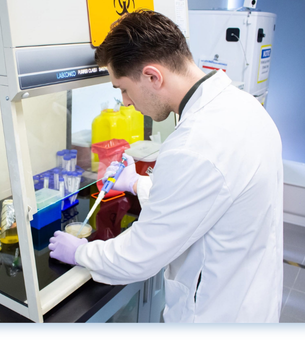
TAV is more directed to decoding how the filament behaves on a physical and molecular scale. The task of student researchers is to design and execute experiments to deconstruct the diffusion kinetics between the polymer, the diffusion surface, and the antibiotic.
Under the guidance of their head of research, Nima Nateghi, and supported by the TAV laboratory supervisor, Elie Saade, the students involved get real, hands-on experience in both foundational and translational research. Three of the first students to work on the project were featured in the scientific article, “A Nanoporous 3D-Printed Scaffold for Local Antibiotic Delivery”, which established preliminary diffusion observations of both polymers, even though the original goal was to develop 3D-printed bone cement beads.
“Having research experience up your sleeve puts you at a huge advantage when you get to university.” David I. (student researcher, 2024)
What we have done
So far, the TAV laboratory has explored how different printing parameters affect diffusion, experimented with dyes as simulated antibiotics, and established multiple experimental designs and analysis protocols to reverse-engineer the diffusion kinetics of both filaments.
In 2021, the lab looked into the fundamental behaviour of both filaments by loading disks of both materials with antibiotics and placing them on bacteria. This is called a Kirby-Bauer assay, which allows us to have a visual indicator of how an antibiotic diffuses out in the shape of a ring around the disk. Their work confirmed there was statistically no difference in the diffusion pattern between LAY-FOMM and LAY-FELT; furthermore, while both materials diffused at the same rate as the commercial disk within the first 24 hours, they showed a slower release later on than expected, indicating it takes longer for the antibiotic to move within the 3D-printed disks than the commercial control disks.
The following years have been dedicated to explaining this phenomenon. Students from 2022 and 2024 focused attention on hydrogel comparison and dye simulations, as well as experimenting with disk surface-to-volume ratios. Within the past year, while no publishable results were collected, TAV students solidified multiple techniques, experimental plans, and disk parameters for the next generation of students to build upon.
“I enjoyed learning how to design controlled experiments, analyze diffusion data, and feel a part of something meaningful.” -Alana C. (student researcher, 2025/26)
What comes next?
During the summer of 2026, there were multiple issues regarding the printing process. Due to the filament having such unique properties, the printers have a hard time printing the polymer at high precision as they do plastic filaments. This, in turn, caused a lack of disks to use for the different experiments planned. While the printer was being troubleshooted, the direction of the project was shifting towards experimental design involving diffusion on real human cells as well as animal wounds. While not much was set in motion, the groundwork was laid neatly in hopes of the next team of students doing both kinetic decoupling and live tissue experiments concurrently.
References
Ahangar, P., Akoury, E., Ramirez Garcia Luna, A. S., Nour, A., Weber, M. H., & Rosenzweig, D. H. (2018). Nanoporous 3D-Printed Scaffolds for Local Doxorubicin Delivery in Bone Metastases Secondary to Prostate Cancer. Materials, 11(9), 1485. https://doi.org/10.3390/ma11091485
Ahangar, P., Li, J., Nkindi, L. S., Mohammadrezaee, Z., Cooke, M. E., Martineau, P. A., Weber, M. H., Saade, E., Nateghi, N., & Rosenzweig, D. H. (2024). A Nanoporous 3D-Printed Scaffold for Local Antibiotic Delivery. Micromachines, 15(1), 83. https://doi.org/10.3390/mi15010083
Cooke, M. E., Ramirez-GarciaLuna, J. L., Rangel-Berridi, K., Park, H., Nazhat, S. N., Weber, M. H., Henderson, J. E., & Rosenzweig, D. H. (2020). 3D Printed Polyurethane Scaffolds for the Repair of Bone Defects. Frontiers in Bioengineering and Biotechnology, 8. https://doi.org/10.3389/fbioe.2020.557215
3D-Printed Nanoporous Scaffolds Impregnated with Zoledronate for the Treatment of Spinal Bone Metastases. (2026). ResearchGate. https://doi.org/10.1557/adv.2019.156
You, J., Preen, R. J., Bull, L., Greenman, J., & Ieropoulos, I. (2017). 3D printed components of microbial fuel cells: Towards monolithic microbial fuel cell fabrication using additive layer manufacturing. Sustainable Energy Technologies and Assessments, 19, 94–101. https://doi.org/10.1016/j.seta.2016.11.006